Spatial Profiling, Analytical Approaches, and New Transcriptomic Technologies
Spatial profiling approaches are continuously advancing as researchers progressively break new ground in imaging techniques at greater resolutions. When running spatial analysis of cellular behaviour, maintaining the context of the cell in surrounding tissue is essential for developing an understanding of cell signalling and cellular interactions. Advances in single-cell imaging have facilitated a huge leap in understanding regarding the behaviour of individual cells.
However, studies of cell signalling – relevant to investigating cancers and the tumour microenvironment (TME) – need to be carried out through multi-omics analysis. Two presentations at Spatial Biology US: 2022 explored current and future approaches to fully-resolved spatial imaging, with consideration given to how their applications could shape future investigations into the behaviour and function of the TME. These approaches include the computational package Giotto, and the barcoding method Slide-seq.
Spatial Profiling: Interpreting the Spatial Environment with Transcriptomics
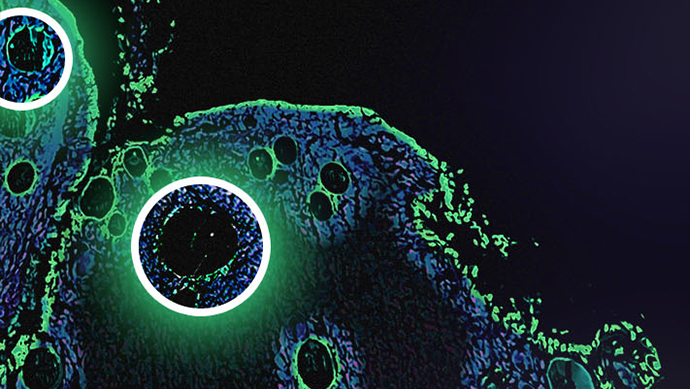
At present, a major focus in the spatial imaging community is on the use of transcriptomics and other approaches to interpret the spatial environment. Being able to resolve the tissue environment at multiple scales is integral to gaining additional context on how cancers interact with tissue, and can provide insights on the efficacy of a particular treatment approach. One avenue for in situ transcriptome imaging involves the use of sequential fluorescence in situ hybridization (seqFISH+), which can image the mRNA of tens of thousands of genes in single cells with a high degree of accuracy.
Guo-Cheng Yuan, Professor of Computational Biology at the Icahn School of Medicine at Mount Sinai, expanded on how spatial profiling approaches can be used for comprehensive investigations of cellular data – with or without the assistance of computational tools. “The spatial patterns can go all the way from the cell or sub-cell level down to these tissue levels,” he explained. “No single method can address all of this, so to help we’ve been trying to develop a general affordable, comprehensive pipeline.”
Applications of Giotto for Interpreting Spatial Transcriptomics Data
The pipeline Yuan has been developing is Giotto: a package which provides the tools to process, analyse, and visualise single-cell spatial expression data. The justification behind developing Giotto came from a common issue with other analytical approaches. Most spatial data does not arrive at a single-cell resolution, which poses issues for interpreting the associated tissue information. Yuan explained that his team had set out to make a technology with broad opportunities for application focusing on the data structure, rather than the underlying raw data itself. Giotto is applicable for analysing data from either sequencing-based or imaging-based platforms.
- Celebrating the achievements of Barbara McClintock, pioneer of gene transposition
- New human pangenome capturing more genetic diversity released
- Advanced data analysis pipelines for spatial transcriptomics
A key focus for investigation through Giotto was the analysis of spatial contexts for different spatially-identified genes. “While we look at the data, we are not just looking at the gene expression pattern of the cell itself but also the process as well,” said Yuan. He added that the software enabled a variety of interrogative approaches, including cell analysis, deconvolution, and cell interaction analysis. The spatial transcriptomic analysis enabled through this approach is a powerful tool for cancer research, facilitating the systemic characterisation of the TME and allowing the identification of cell-cell interaction mechanisms.
Retaining Tissue Contexts in Genomic Measurements
Spatially-resolved approaches have enabled something close to a genomics revolution in recent years. Fei Chen, Assistant Professor of Stem Cell and Regenerative Biology at Harvard, and a Core Institute Member of the Broad Institute of Harvard and MIT, explained his excitement about the prospect of measuring the world of genomics and the world of microscopy. “We’re able to identify cell states using cell sequencing and single cells, and we’ve begun to find their regulatory mechanisms and molecular identities.” However, these investigations are often carried out on dissociated tissues, making it difficult for researchers to follow up the meaning of the cell states identified in the absence of tissue contexts.
As such, a central focus in this avenue of research has been the rapid development of imaging tools and spatial genomic tools, as well as their convergent use. “We would like to place those cells in the context of tissue and understand their interactions with each other,” said Chen, giving the example of hundreds of thousands of cells from a mouse brain. To help progress an understanding of cellular signalling pathways, researchers would benefit from being able to understand the relationships of those cells and the cellular niches they live in.
“We’re able to identify cell states using cell sequencing... and we’ve begun to find their regulatory mechanisms and molecular identities.”
Another focus in spatial profiling is the amount of variance in gene expression that can be conferred from a cell’s spatial context. “We see a lot of variability in single cell data, and these are just intrinsic biological points,” Chen explained. One future aim for spatial imaging could be discovering cell type-specific spatial gene expression patterns that explain the gene expression of the cell.
High-Throughput, Genome-Wide Readouts of Gene Expression with Slide-seq
While the spatial positions of cells in tissue strongly influence function, a high-throughput, genome-wide readout of gene expression with cellular resolution is lacking. One approach to circumvent this is Slide-seq: a method developed by Chen and his team for transferring RNA from tissue sections onto a surface covered in DNA-barcoded beads with known positions. It functions as a micron bead array with sequenced bead barcodes. Each bead contains millions of oligonucleotides, and is used for capturing mRNA.
“When we run the experiment, the RNA is locally captured onto the tissue,” explained Chen. “Since you already have the spatial locations for each one of these barcodes, you can match them up, and now you have a spatial location for each 10-micron read.” Information conveyed by Slide-seq allows the visualisation and recreation of cellular processes at high resolution. Chen explained that his team had developed a model for taking a set of spatial transcriptomic data and then using unsupervised learning approaches to assign a proportion of cell types to a particular area. This information is then deconvoluted for interpretation.
As Chen explained, the main purpose of spatial transcriptomics in this context is to understand the spatial environment through looking at cell-type effects on gene expression. “You get this capture process, and there are different capture processes that form part of this expression,” said Chen.
Investigative Approaches for Spatial Imaging: Tomorrow, Today
Advances in spatial transcriptomics approaches such as the ones explored in this article have facilitated the development of incisive techniques for uncovering new information about the cellular microenvironment. Giotto and Slide-seq are two techniques on the cutting edge of spatial profiling and investigation, and the potential for their application in furthering understandings of cellular interactions and gene expression is enormous.
Want to keep up with the latest developments in spatial transcriptomics and imaging? Sign up to our Omics newsletter to receive a monthly summary of industry trends and new advances in the field straight to your inbox. If you’d like to know more about our upcoming Spatial Biology Europe conference, visit our event website to download an agenda and register your interest.