Decoding the Language of Tumor Immune Microenvironment: A Deep Dive into Spatial Biology Techniques
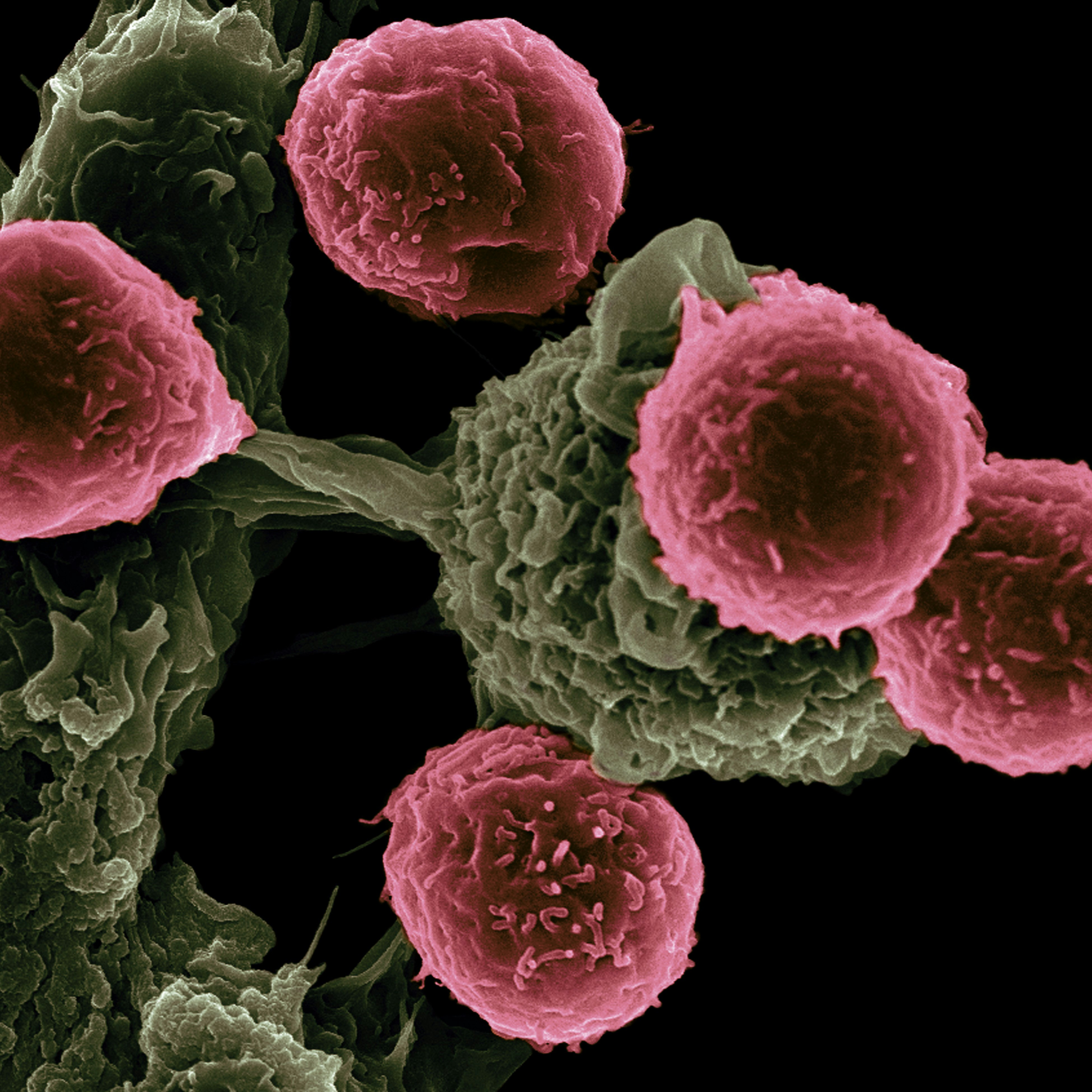
The tumor immune microenvironment (TIME) is a complex network of interactions between tumor cells and various components of the immune system. Understanding the dynamic nature of this microenvironment is crucial for developing effective cancer therapies. The spatial organization of cells within the tumor microenvironment plays a critical role in determining the immune response and the success of immunotherapy. Immuno by Oxford Global will delve into the fascinating world of spatial biology techniques and their role in decoding the language of the tumour immune microenvironment.
Understanding the Role of Spatial Biology in Tumor Microenvironment
Spatial biology, aims at decoding the spatial arrangements and interplays of cells within tissue environments. In the specific context of the tumor microenvironment, spatial biology techniques serve as indispensable tools, offering nuanced insights into the precise localization and distribution of various cell types, including immune infiltrates, stromal cells, and tumor cells, along with their dynamic functional states. Through advanced imaging modalities and spatial profiling approaches, researchers can decipher the spatial relationships between these heterogeneous cell populations, unveiling critical insights into the spatially orchestrated immune responses to cancer, thus identifying potential targets for therapy and advancing precision medicine strategies aimed at improving cancer treatment outcomes.
Key Components of the Tumor Immune Microenvironment
The tumor immune microenvironment represents a highly intricate ecosystem characterized by a myriad of interacting cellular components, signalling molecules, and extracellular matrix elements. At its core are a diverse array of players, including tumor cells, immune cells encompassing T cells, B cells, and natural killer cells, stromal cells like fibroblasts and endothelial cells, as well as immune checkpoints such as PD-L1 and CTLA-4. Each of these constituents contributes uniquely to the complex interplay within the tumor microenvironment, orchestrating the immune response and influencing tumor progression. Spatial biology techniques emerge as indispensable tools in this landscape, offering researchers a lens to peer into the intricate spatial organization and quantify the abundance of these components. Through advanced imaging modalities and spatial profiling approaches, researchers gain the ability to dissect the spatial relationships and distributions of these entities, thus unraveling their functional significance and potential therapeutic targets within the tumor microenvironment.
Techniques for Analysing Spatial Biology in Tumor Microenvironment
- Immunohistochemistry
Immunohistochemistry (IHC) is a widely used technique in spatial biology that allows researchers to visualize specific proteins within tissue sections. By using antibodies that bind to target proteins, researchers can identify and quantify the expression levels of immune cell markers, immune checkpoints, and other proteins of interest. IHC provides information on the localization and abundance of these proteins, enabling researchers to decipher the interactions between tumour cells and immune cells in the microenvironment.
- In situ Hybridization
In situ hybridization (ISH) is a technique used to detect and localize specific RNA molecules within tissue sections. By using complementary nucleic acid probes, researchers can identify and visualize the expression of genes involved in immune responses, such as cytokines, chemokines, and immune cell markers. ISH provides valuable information on the spatial distribution of gene expression within the tumour microenvironment, shedding light on the communication between tumour cells and immune cells.
- Spatial Transcriptomics
Spatial transcriptomics is a cutting-edge technique that combines spatial information and gene expression profiling. It allows researchers to simultaneously visualize the spatial distribution of RNA molecules and measure their expression levels within tissue sections. By mapping the transcriptome of the tumor microenvironment, spatial transcriptomics provides a comprehensive view of the gene expression patterns and cellular interactions within the microenvironment. This technique has revolutionized our understanding of the immune response in cancer and has the potential to uncover novel therapeutic targets.
- Spatial Proteomics
Spatial proteomics is a powerful technique that enables researchers to study the spatial organization of proteins within tissues. By using mass spectrometry-based approaches, researchers can identify and quantify proteins in specific regions of interest. Spatial proteomics provides insights into the protein-protein interactions and signaling pathways that regulate immune responses within the tumor microenvironment. This technique holds great promise for discovering new targets for immunotherapy and personalized cancer treatment.
Combining Spatial Biology Techniques for Comprehensive Analysis of Tumor Immune Microenvironment
To gain a comprehensive understanding of the tumor immune microenvironment, researchers often combine multiple spatial biology techniques. By integrating data from different techniques, researchers can obtain a more detailed and nuanced view of the interactions between tumor cells and immune cells. For example, combining IHC with spatial transcriptomics can reveal the spatial localization of immune cell markers and their gene expression profiles simultaneously. These multi-dimensional datasets provide a deeper understanding of the cellular dynamics and functional states within the tumor immune microenvironment.
Conclusion and Future Directions in Spatial Biology Techniques for Tumor Immune Microenvironment
Spatial biology, driven by advanced methodologies like multiplexed spatial profiling and single-cell spatial genomics, offers profound insights into the tumor immune microenvironment. These techniques unravel immune cell diversity, interactions, and responses with unprecedented precision, promising enhanced cancer therapies and patient outcomes. When combined with established techniques like immunohistochemistry and spatial transcriptomics, spatial biology provides a comprehensive understanding of cellular dynamics within the microenvironment, paving the way for more effective cancer treatments. The field is rapidly evolving, fueled by breakthroughs in technologies such as multiplexed spatial profiling, single-cell spatial genomics, and spatially resolved proteomics. These advancements enable researchers to explore immune cell diversity, interactions, and responses in the tumor microenvironment with unparalleled precision. As spatial biology techniques continue to advance, they hold significant promise for enhancing cancer therapies and improving patient outcomes.
