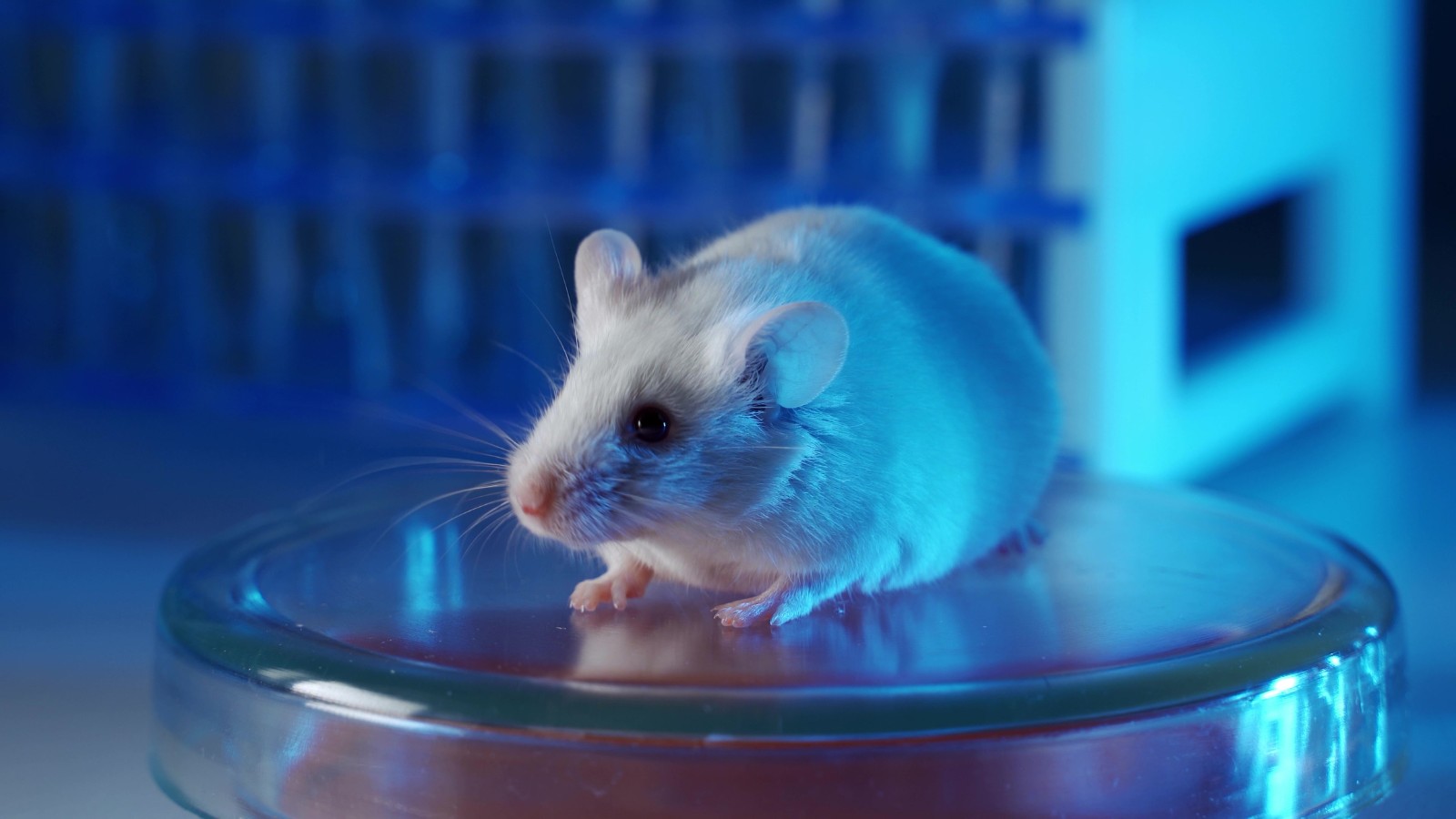
Using Mouse Models for Preclinical Immuno Oncology
Oxford Global’s symposium, Preclinical Immuno-Oncology: Online, featured two presentations from expert scientists which discussed the use of mouse models for preclinical immuno-oncology projects. Here we take a look at these projects and see what can be learnt for the process of preclinical in vivo testing.
Predicting The Safety And Efficacy Of Mouse Models
Stephanie Casey Parks is a Senior Scientist in the department of Oncology at Amgen. One aspect of her research aims to use humanised mouse models to drive next generation cellular therapies, which is what she showcased in her presentation.
Engineered T cell therapy is a rapidly advancing field; however there is still an unmet need present in several cancer indications. Furthermore, the field also faces challenges such as low retention of transfer T cells, loss of tumour antigens, and escape of the residual cells. One way that researchers are working to counter these issues is by adding additional genes that they call ‘payloads’ to TCR-T cells in order to improve their behaviour.
Protein X
Based both on mRNA and protein expression, the target of the TCR-T cells, ‘protein X’, has extremely restricted expression on normal tissue but high expression on many tumour types. Furthermore, high expression of protein X is associated with poor clinical prognosis.
TCR-T cells Targeting Protein X
Dr Casey Parks’ team tried a variety of TCR-T cells against tumour cells with high, medium, and low expression of protein X. The team ended up narrowing in on a TCR-T termed ‘construct A’ which appeared to have the best potency, as well as the cell line B-CPAP, which had the highest expression of protein X. They selected both of these tools to move forward with for their in vivo assays.
In vivo Modelling
Moving forward with their in vivo modelling, the team set up an immunocompromised mouse model to allow for the take of a human xenograft, as well as engraftment of their transferred T cells. Included in the model were TCR-T cells with construct A and the B-CPAP thyroid tumour cell line xenograft.
Their in vivo modelling was conducted using two different ‘flavours’ of models: ‘admix’, where TCR-T cells and tumour cells were mixed in an Eppendorf tube prior to implantation, and ‘established’, where the TCR-T cells were administered in vivo once the tumours had grown to a certain point (100 mm3).
First In vivo Experiment
The first in vivo experiment tested several cell numbers of TCR-T cells with both established and admix tumours. Early results from the established experiment didn’t show any efficacy, but they continued to optimise the model as they saw some differences when escalating dose in the admix experiment.
Cytokine Support
In the interest of improving their results, the team turned to using cytokine support in their modelling, which is in line with how clinical testing would operate. They hypothesised that IL-2 cytokine support would lead to further expansion and persistence of their cells.
This turned out to be very successful. With the cytokine support, the team began to see signs of slowing tumour growth in the established group, while the admix mixture demonstrated complete clearance of the tumours.
Efficacy Study
Moving into an efficacy study, the team were very encouraged to see that TCR-T cells with payload Y showed signs of inhibiting tumour growth. However, tolerability issues were observed in the mice, which meant that the team had to terminate the study.
Optimising the Model
In the interest of continuing to optimise the model, the team hypothesised that they needed to improve the purity of the transferred T cells. They also wanted to test whether a slower-growing tumour model would give them a more suitable runway. Therefore, they pivoted to an NCI-H2023 cell line and titrated in the IL-2.
Data from the experiments on the optimised model showed that ten out of the ten mice that were given TCR-T cells targeting protein X showed robust tumour growth inhibition. Following the animals out after the treatment, a handful of the tumours did begin to grow again, however those tumours remained small, and the end survival rate was 100% by the end of the study. Furthermore, no tolerability issues were observed.
Further research is ongoing to understand cell expansion and persistence, cell homing, and a combination of these T cells with other agents to improve tumour outgrowth control and relapse prevention.
RELATED:
- Delivering Immuno-Oncology Therapies for Solid Tumours
- Improving Preclinical Modelling for IO
- Gene Knockout Mice in Disease Modelling
Human Knock-In Mice For Selecting Next Generation Immune Checkpoint Inhibitors
The second speaker at the symposium was delivered by Thierry Guillaudeux, Chief Scientific Officer at Kineta. His presentation focussed on selecting the most appropriate human knock-in mouse model for their project, an anti-VISTA antibody. Furthermore, he discussed tumour models and analysis for immunological response and the use of human knock-in mice for PK analysis.
The Project
VISTA, an immuno-modulatory molecule, is a member of the B7 family and is closely related to PD-L1. It has a unique expression on myeloid cells as well as a weaker expression on T and NK cells. It counters T cell activation and suppresses T effector cell function.
In tumours, VISTA is a key driver of immunosuppression, it is highly expressed, and associated with poor prognoses in patients. These factors have made it an attractive immuno-oncology target. Therefore anti-VISTA antibodies can block VISTA to drive an immune response against tumour cells.
In vitro
As a proof of concept, the tumour models that Dr Guillaudeux’s team used to test this mechanism were a CT26 (colon carcinoma) and a B16F10 tumour model. As a single agent, they saw moderate efficacy of an anti-VISTA antibody, but significant improvement when combined with immune-checkpoint inhibitors
Using what they had learnt, the team began working on a screening process using over 100 fully human antibodies to find the best lead candidate. For this, they looked for inhibition of MDSCs, activation of monocytes, and the enhanced activation of T cells.
Using Human Knock-In Mice
The team then went looking for a human knock-in mouse model to recapitulate their experiments in vivo. The model that they selected was a human VISTA knock-in mouse model produced by Genoway.
They were particularly interested in the VISTA expression in the myeloid compartment of the CD11b+ cells in these knock-in mice as that was the target that they were focussing on. Testing this in the lab confirmed a desirable expression of human VISTA and an absence of mouse VISTA compared to the wild type mice.
Testing the Lead Antibody
Focusing on cold tumours, they evaluated the efficacy of Kineta’s anti-VISTA lead antibody. First, they tested their lead on the cold tumour MC38 (colon carcinoma). Here, they witnessed a positive response to the lead both as a single agent and combined with anti-PD-1.
Next, they evaluated against another cold tumour, MB49 (bladder cancer). This showed very strong single agent activity. Analysing the tumour microenvironment of the mouse, they saw an increase in both NK and T cells infiltrating the tumour. In a more inflamed tumour, EG-7 (T cell lymphoma), there was also strong single agent activity
PK Analysis
Finally, the team used their mouse models to perform PK analysis on their lead candidates. Dr Guillaudeux said that analyzing their lead antibodies in VISTA knock-in mice allowed them to understand their candidates much better and aided in lead selection.
Join our two-day Immuno event packed with leading speakers, research and cutting-edge presentations on the latest advancements in cancer immunotherapy, with dedicated sessions o n different therapeutic modalities as well as case studies on innovative preclinical and translational tools.